Somatosensory Psychophysics
Adapted from MS1 Neuroscience Course, 1993
Dr. Joel D. Greenspan
Objective:
To explain in short essays or diagrams the anatomical and/or physiological basis for the distribution of somatosensory cold and warm spots and the regional variation of tactile acuity, at the level of 85% proficiency for each student.
In order to achieve this objective, you will need to be able to:
- Examine the distribution of cold and warm sensitive spots on the skin.
- Examine tactile acuity of various areas of skin
Materials
Group Supplies:
Stainless Steel probes in ice
6 inch plastic ruler
2 paper clips
Thermal sensitivity
Methods
For this laboratory, you will work in groups of three. Each one of you will take turns being the experimenter, the subject, and the record keeper. Each person, then, will have his/her own laboratory data sheets to turn in at the end.
We will begin this laboratory with a demonstration of the discrete nature of cutaneous sensitivity. If you use a stimulating probe that is small enough, you can determine spots on your skin that are particularly sensitive, and other spots that are less so. This phenomenon is most apparent with your sense of coolness, which we will examine today.
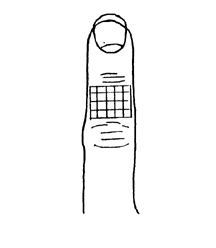
You will be using a stainless steel probe to serve as your stimulator. Since the pen tip is a good heat sink, and is cooler than your skin, it provides an adequate cooling stimulus. The subject should be seated, and not able to see the stimulus application. Usually, the subject would be blindfolded, but in this case, we’ll count on the subject keeping his/her eyes closed. The subject will place his/her left hand on the table, palm down. The experimenter will envision a twenty grid array on the middle phalange of digit three (see figure in lab data sheet). If digit three is injured, use another digit or the right hand. The experimenter will lightly touch the subject’s finger at each of these twenty spots. He/she should maintain contact with the skin for 2-3 seconds (enough contact to indent the skin a slight amount), and then remove the pen. Then, the subject will say whether he/she felt a cool sensation or not, and the record keeper will write down the response. The experimenter should wait 5-6 seconds before the next stimulus. You may wish to practice at a few points first before collecting the data. At the end, you will have a map of cool spots, as well as a count of how many of the 20 spots felt cool. We will look at the class distribution of cool spot density in class.
Results
COOL SPOT MAPPING
Indicate with a filled circle which spots provided a sensation of coolness, and use a hollow circle for spots that were otherwise.
|
Total # cool spots |
/20 |
Tactile (two-point) discrimination
Methods
For this laboratory, you will work in groups of three. Each one of you will take turns being the experimenter, the subject, and the record keeper. Each person, then, will have his/her own laboratory data sheets to turn in at the end.
The class will perform a set of experiments to evaluate one measure of tactile acuity: two-point discrimination. As before, you will work in groups of three, each taking turns at being the subject, the experimenter, and the record keeper.
The basic question that you will be asking is, “How much of a separation between two points is needed in order to perceive them as two distinct points?” The minimal separation that is needed for perceiving two distinct points is called the two-point threshold. You will determine the two-point threshold on different sites of your hand, in order to demonstrate the extent of regional variability.
The protocol that you will be using is the method of limits. For the sake of time, you will perform one ascending series of stimuli (e.g., progressively larger point separations), and one descending series of stimuli for each skin site tested. Normally, one would repeat these series several times and determine an average.
The subject should be seated, with his/her eyes closed. To begin an ascending series, the experimenter sets the two-point device so that the two probes are as close as possible. The experimenter applies the stimulus by pushing the probes down on the desired skin site for 2-3 sec. The subject then reports whether he/she felt “one” or “two” points. As long as the subject reports “one”, the experimenter increases the distance between the two points from stimulus to stimulus, being careful to use a consistent amount of force each time. When the subject gives a response of “two”, the ascending series is over, and the record keeper notes the separation that was used to evoke the response of “two”. Technically, the record keeper should use the response of “two”, and the previous separation that was perceived as “one”. Thus, if you were increasing the separation in l-mm steps (ascending series), and the first response of “two” was given at a separation of 8mm, then the threshold value of that series would be 7Smm (since the previous stimulus of 7mm yielded a response of “one”). To begin the descending series of stimuli, adjust the two point device so that it has a separation several millimeters greater than the threshold determined by the ascending series. Then, the experimenter repeats the steps described above, only this time the separation is reduced each time the subject reports perceived as “one”.
You will then take the average of the ascending and the descending series thresholds to determine that subject’s two-point threshold for that skin site.
You will be testing four sites on each subject’s right hand (palmar surface). Site # l is the tip of digit 2; site # 2 is the proximal half of the distal phalanx; site # 3 is the middle of the proximal phalanx of digit 2; site # 4 is the middle of the thenar eminence (muscle at the base of the thumb). If a person has an injury on digit 2, another digit should be used.
You will also be testing for sites on each subjects. Site # 5 is the right hand (posterior surface), site # 6 is the right shoulder (deltoid region), site # 7 is the upper back (interscapular), site # 8 is the right cheek (zygomatic region), site # 9 is the right knee (anterior), and site # 10 is the right lower leg (posterior crual). Group results will be tabulated.
Results
TWO-POINT DISCRIMINATION
|
Site # 1 |
Tip of digit # 2, right hand |
|
Ascending series threshold |
- mm |
|
Descending series threshold |
- mm |
|
Average two-point threshold |
- mm |
|
Site # 2 |
Proximal
half of distal phalanx |
|
Ascending series threshold |
- mm |
|
Descending series threshold |
- mm |
|
Average two-point threshold |
- mm |
|
Site # 3 |
Middle of proximal phalanx |
|
Ascending series threshold |
- mm |
|
Descending series threshold |
- mm |
|
Average two-point threshold |
- mm |
|
Site # 4 |
Thenar eminence, right hand |
|
Ascending series threshold |
- mm |
|
Descending series threshold |
- mm |
|
Average two-point threshold |
- mm |
|
Site #5 |
Interscapular region, upper back. |
|
Ascending series threshold |
- mm |
|
Descending series threshold |
- mm |
|
Average two-point threshold |
- mm |
|
Site # 6 |
Zygomatic region, right cheek |
|
Ascending series threshold |
- mm |
|
Descending series threshold |
- mm |
|
Average two-point threshold |
- mm |
Discussion
1. Explain the anatomical and/or physiological basis of the distribution of the cold and warm spots..
2. What were you able to determine about regional variation of tactile acuity?
3. What sources of error could affect the results of these experiments?
4. What procedural modifications could reduce or eliminate such errors?
5. What neuroanatomical and neurophysiological features determine two-point threshold?
© David G. Ward, Ph.D. Last modified by wardd 23 May, 2006