Models of Chemical Messengers
Objective:
To construct using models or diagrams the organization of channel linked receptors, enzyme linked receptors, and G-protein linked receptors, and to explain in short essays or flow charts their role in cellular communication, at the level of 85% proficiency for each student.
In order to achieve this objective, you will need to be able to:
- Construct a model of a cell membrane.
- Construct models of channel linked receptors.
- Diagram the behavior of channel linked receptors and the action of Ca++ and protein kinases.
- Construct models of enzyme linked receptors.
- Diagram the behavior of enzyme linked receptors and the action of kinases.
- Construct models of G-protein linked receptors.
- Diagram the behavior of G-protein linked receptors and the action of cyclases, cAMP, cGMP, and protein kinases.
- Diagram the action of phospholipases, IP3 and Ca++, DAG and protein kinases.
Materials:
Group Supplies
Computer with the Chime plug-in installed for Internet Explorer
(The Chime plug-in has been pre-installed onto the computers in the physiology lab)
Structures of lipid bilayer
http://molvis.sdsc.edu/atlas/atlas.htm
(These files have been downloaded and pre-loaded onto the computers in the physiology lab)
Structures of membrane proteins
http://blanco.biomol.uci.edu/Membrane_Proteins_xtal.html
(These files have been downloaded and pre-loaded onto the computers in the physiology lab)
Phospholipid bilayer
Cotton swabs (phospholipids for cell membrane)
Channel linked receptors
White
clay (a chemical messenger)
Drinking straws (channel linked receptors)
Colored beads (various ions)
White beads (calcium ions)
¼ Popsicle stick with White clay (calmodulin)
¼ Popsicle stick with Purple clay (protein kinase)
Orange
beads (inorganic phosphate)
Red clay (a protein)
Enzyme linked receptors
White
clay (a chemical messenger)
Glue stick with a purple end (enzyme linked receptor with tyrosine kinase)
Glue stick with a green end (enzyme linked receptor with guanylate cyclase)
Green clay (guanine nucleotide)
¼ Popsicle stick with Purple clay (protein kinase)
Orange
beads (inorganic phosphate)
Red clay (a protein)
G-protein linked receptors
White
clay (a chemical messenger)
Glue sticks (G-protein linked receptors)
Yellow clay (G-protein)
Green clay (guanine nucleotide)
Blue clay (adenine nucleotide)
¼ Popsicle stick with green clay (guanylate cyclase)
¼ Popsicle stick with blue clay (adenylate cyclase)
¼ Popsicle stick with Purple clay (protein kinase)
Orange beads (inorganic phosphate)
Red clay (a protein)
Yellow clay (G-protein)
Green clay (guanine nucleotide)
Orange beads (inorganic phosphate)
¼ Popsicle stick with orange clay (phospholipase)
Cotton swab (phosphatidyl Inositol)
Cotton swab shortened, with orange clay (IP3)
White beads (calcium ions)
Cotton swab shortened, with gray clay (DAG)
¼ Popsicle stick with Purple clay (protein kinase)
Orange beads (inorganic phosphate)
Red clay (a protein)
Methods:
Cell Membrane
Double click the shortcut named lipid bilayer to examine the three dimensional organization of cel membrane structures:

In order to examine the three dimensional organization of the lipid bilayer, pick Lipid Bilayer from the list on the computer; for example:
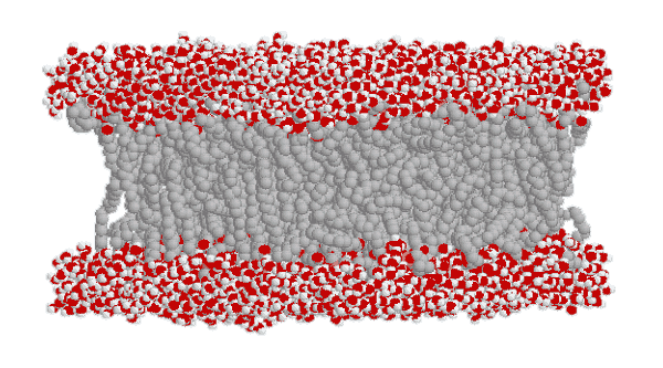
Based on what you see, construct a section of a cell membrane using 20 cotton swabs to represent the phospholipids of the lipid bilayer
Channel Linked Receptors
In order to examine the organization of a transmembrane channel, pick Aquaporin from the list on the computer; for example:
Note that this water channel is very complex. Ion channels are just as complex. We will simply use a length of drinking straw to represent a channel. Place two channels (drinking straws) through the lipid bilayer you previously constructed.
Bind a chemical messenger to a receptor linked channel and use clay and beads to construct the sequence of events leading to the movement of ions through an open channel. Use the following table as a guide. Other ions may be explored.
|
Ca++ directly changes membrane potential |
|
|
overall action |
specific steps |
|
Ca++ channel activation |
A chemical messenger binds to the Ca++ channel |
|
Ca++ makes the intracellular fluid more positively charged |
Ca++ flows through the channel into the cytoplasm |
Bind a chemical messenger to another receptor linked channel and use beads and clay to construct the sequence of events leading to indirect changes in the cell via second messengers. Use the following table as a guide.
|
Ca++ acts as a secondary messenger |
|
|
overall action |
specific steps |
|
Ca++ channel activation |
A chemical messenger binds to the Ca++ channel |
|
Ca++ flows through the channel into the cytoplasm |
|
|
Ca++ change the activity of an intracellular protein
|
Ca++ binds to Calmodulin |
|
Calmodulin in turn activates a Protein Kinase |
|
|
The Protein Kinase in turn phosphorylates a Protein |
|
Enzyme Linked Receptors
Enzyme linked receptors are just as complex. We will simply use glue sticks to represent enzyme linked receptors. Place two glue sticks through the lipid bilayer you previously constructed.
Bind a chemical messenger to the enzyme linked receptor and use clay, beads and Popsicle sticks to construct the sequence of events leading to cellular changes via second messengers. Use the following table as a guide
|
Enzyme linked receptors act through second messengers |
|
|
enzyme |
specific steps |
|
|
|
|
Tyrosine Kinases: |
Receptor activation: |
|
Tyrosine Kinases phosphorylate tyrosine in target proteins |
|
G-protein Linked Receptors involving a cyclase
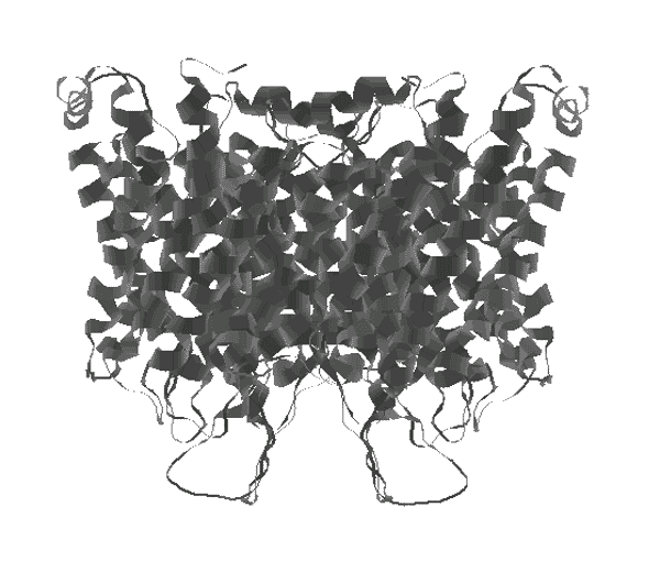
In order to examine the three dimensional organization of a single G-protein coupled receptor, pick G-protein coupled receptor from the list on the computer. For example:

Note that G-protein linked receptors are also very complex. We will again use a length of glue stick to represent a G-protein linked receptor. Place two G-protein linked receptors through the lipid bilayer you previously constructed.
Bind a chemical messenger to the G-protein linked receptor and construct using clay, beads and Popsicle sticks, the sequence of events leading to cellular changes via a cyclase and second messengers. Use the following table as a guide.
|
G-protein linked receptors act through a cyclase |
|
|
enzyme |
specific steps |
|
|
|
|
Adenylate Cyclase |
G-protein is bound to GDP |
|
Receptor activation: |
|
|
G-protein releases GDP and binds GTP |
|
|
G-protein activates Adenylate Cyclase |
|
|
Adenylate Cyclase catalyses ATP to cAMP |
|
|
cAMP activates Protein Kinase A |
|
|
Protein Kinase A phosphorylates a protein |
|
|
cAMP degraded by cAMP phosphodiesterase (inhibited by caffeine) |
|
|
|
|
|
Guanylate Cyclase |
G-protein is bound to GDP |
|
Receptor activation: |
|
|
G-protein releases GDP and binds GTP |
|
|
G-protein activates Guanylate Cyclase |
|
|
Guanylate Cyclase catalyses GTP to cGMP |
|
|
cGMP activates a Protein Kinase G |
|
|
Protein Kinase G phosphorylates a Protein |
|
|
cGMP degraded by cGMP phosphodiesterase (inhibited by caffeine) |
|
G-protein Linked Receptors involving a phospholipase
We will again use a length of glue stick to represent a G-protein linked receptor. Place two G-protein linked receptors through the lipid bilayer you previously constructed.
Bind a chemical messenger to the G-protein linked receptor and construct using clay, beads and Popsicle sticks, the sequence of events leading to cellular changes via phospholipase and second messengers. Use the following table as a guide.
|
G-protein linked receptors act through a phospholipase |
|
|
enzyme |
specific steps |
|
|
|
|
Phospholipase C |
G-protein is bound to GDP |
|
Receptor activation: |
|
|
G-protein releases GDP and binds GTP |
|
|
G-protein activates Phospholipase C |
|
|
Phospholipase C catalyses Phosphatidylinositol to Inositoltriphosphate (IP3) |
|
|
IP3 opens Ca++ channels in Endoplasmic reticulum |
|
|
|
|
|
Phospholipase C |
G-protein is bound to GDP |
|
Receptor
activation: |
|
|
G-protein releases GDP and binds GTP |
|
|
G-protein activates Phospholipase C |
|
|
Phospholipase C catalyses
Phosphatidylinositol to |
|
|
DAG activates Protein Kinase C |
|
|
Protein Kinase C phosphorylates a protein |
|
Discussion:
1. On which side of the cell membrane is the receptor located for the channel linked receptors, the enzyme linked receptors, and the G-protein linked receptors?
2. What sort of chemical messengers bind to these receptors?
3. Diagram the behavior of channel linked receptors and the action of Ca++ and protein kinases.
4. Diagram the behavior of enzyme linked receptors and the action of cyclases and cGMP.
5. Diagram the behavior of G-protein linked receptors and the action of cyclases, cAMP, cGMP, and protein kinases.
6. Diagram the behavior of G-protein linked receptors and the action of phospholipases, IP3 and Ca++, DAG and protein kinases.
© David G. Ward, Ph.D. Last modified by wardd 23 May, 2006