Electromyography and Reflexes
Objective:
To describe in short essays or diagrams the anatomical organization of muscle cells and the neuromuscular junction and to explain how nervous stimulation causes muscle contraction, at the level of 85% proficiency for each student.
In order to achieve this objective, you will need to be able to:
- Describe the electromyographic (EMG) response of muscle during contraction of flexor and extensor muscles.
- Measure changes in the EMG as the force exerted by a muscle increases
- Measure the EMG activity of different muscle groups during movement.
- Measure reaction time of reflexive movements.
Materials
Group Supplies:
self-adhesive, pre-gelled, disposable electrodes
reusable leads
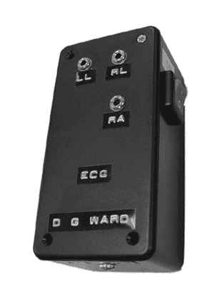
ECG – EMG preamplifier (D. G. Ward) (set to record activity within a range of 30–1,000 Hz., with a gain of 2,000)
digital to analog converter Dataq, DI-154)
computer with display software (WinDaq and WinDaq Browser)
handgrip exerciser
telegraph style switch
percussion hammer modified to send a signal to the computer
Descriptions of stretch reflexes are available as a supplement: Stretch Reflexes
Methods and Results
The construction of the telegraph style switch and the modification to the percussion hammer is carried out as shown below.
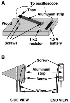
The preamplifier for ECG / EMG is identified as follows. For each set of measurements described below one electrode is plugged into LL and the other electrode is plugged into RA. A third, indifferent electrode, is plugged into RL.
Flexor muscles of the forearm
Flexion
Procedure. Two electrodes are placed diagonally across the flexor muscles of the forearm (Flexor Digitorum and Flexor Carpi groups). The indifferent electrode is placed on the lower body. The arm is held with the elbow by the side of the body and the forearm extended with the fingers curled into a loose fist. The hand is slowly moved toward the front of the wrist (flexion) and then the back of the wrist (extension) to determine for which movement more EMG activity is observes.
Results
|
EMG activity with Flexion |
|
|
EMG activity with Extension |
|
Force and EMG
Procedure. The handgrip exerciser is slowly squeezed, with the computer collecting data and any change in the EMG is noted.
Results
|
EMG activity with light squeezing |
|
|
EMG activity with intermediate squeezing |
|
|
EMG activity with maximal squeezing |
|
Extensor muscles of the forearm
Extension
Procedure. Two electrodes are placed diagonally across the extensor muscles of the forearm (Extensor Digitorum and Extensor Carpi groups). The indifferent electrode is placed on the lower body. The arm is held with the elbow by the side of the body and the forearm extended with the fingers curled into a loose fist. The hand is slowly moved toward the front of the wrist (flexion) and then the back of the wrist (extension) to determine for which movement more EMG activity is observed.
Results
|
EMG activity flexion |
|
|
EMG activity with extension |
|
Temporal Relationship Between EMG and Movement Reaction Time
Procedure. The hand is placed palm down with the forefinger on the switch. The output of the switch goes to one channel on the computer and the EMG to the other channel. With the computer collecting data the subject quickly lifts the forefinger. The students determine whether the EMG change occurs first, the finger movement occurs first, or both occur simultaneously.
Results
|
time between switch and EMG |
|
|
time between switch and movement |
|
|
time between EMG and movement |
|
Reaction Time
Procedure. The EMG and switch outputs are left on separate computer channels, and the hammer is connected to a third channel. The indifferent electrode is placed on the lower body. The subject sits with eyes closed and forefinger on the switch. With the computer collecting data another student strikes the hammer on the table or other surface (using the ‘‘pointed’’ part of the hammerhead, and not the switch), and the subject lifts the finger rapidly on hearing the strike. On the computer, three times are determined: reaction time (hammer strike—when the computer is triggered—to finger lift), the supraspinal response initiation time (hammer strike to start of change in EMG activity), and the electromechanical delay (start of change in EMG activity to finger lift). Students do 10 trials and plot the means 6 SE of reaction time and its components. They then plot a scatter graph of reaction time for the 10 trials each of the two components and calculate correlation coefficients to determine which component is better correlated with reaction time. The present experiment takes advantage of the reaction time variability that tends to occur across trials; no variables are manipulated. The basic question that is addressed is whether this variability is mainly due to processes occurring between the occurrence of the stimulus and the muscle action potentials or between the muscle action potentials and the lifting of the finger. This will be reflected as a greater correlation between that particular component and the total reaction time. Alternatively, both processes could be making equal contributions to the variability in the reaction time, in which case the two correlations will be about the same.
Results
|
trial |
Time between strike and EMG |
Time between strike and movement |
Time between EMG and movement |
|
|
|
|
|
|
|
|
|
|
|
|
|
|
|
|
|
|
|
|
|
|
|
|
|
|
|
|
|
|
|
|
|
|
|
|
|
|
|
|
|
|
|
|
|
|
|
|
|
|
|
mean ± sd |
|
|
|
Quadriceps stretch reflex
Procedure. Two electrodes are placed parallel to the extensor muscles of the thigh (Quadriceps group). The indifferent electrode is placed on the upper body. Students should bring short pants to wear for this experiment. The subject sits with the leg with the electrodes crossed over the other leg, and the experimenter strikes the hammer on the patellar tendon just below the kneecap, triggering the computer and eliciting a knee jerk. Ten trials are given this way and ten more trials are given in which the hammer is struck at a point near the tendon, which does not elicit a reflex; the subject jerks the leg when detecting the tap. For each trial, the time between the stimulus and the EMG change is measured. For the first 10 trials, this represents the spinal reflex latency; for the second 10 trials, it is the supraspinal response initiation time.). The means 6 SE are plotted. one of these time periods is greater than the other, and if so, which one is greater.
Results
|
Strike patellar Tendon |
Strike near Tendon |
||
|
trial |
Time between strike and EMG |
trial |
Time between strike and EMG |
|
|
|
|
|
|
|
|
|
|
|
|
|
|
|
|
|
|
|
|
|
|
|
|
|
|
|
|
|
|
|
|
|
|
|
|
|
|
|
|
|
|
|
|
|
|
|
|
|
|
|
mean ± sd |
|
mean ± sd |
|
Discussion
1. Explain what causes the EMG.
2. Explain the change in EMG activity as the force exerted by a muscle increases.
3. Explain why the EMG activity of extensors and flexors is different during a movement.
4. Explain the timing of the EMG relative to the contraction of a muscle.
5. Explain the time delay seen between intending to make a movement; and the change in EMG activity.
6. Explain the differences in time delay between a stimulus and a reflex EMG response; and the time delay between a stimulus and a voluntary EMG response.
© David G. Ward, Ph.D. Last modified by wardd 23 May, 2006