The Electrocardiogram
Note: This lab may be conducted by itself or integrated with the next lab: The Electrocardiogram and Cardiac Pulse.
Objective:
To explain in a short essay or with a diagram the significance of the P, QRS, and T waves, and to calculate heart rate and wave intervals in an ECG print out, at the level of 85% proficiency for each student.
In order to achieve this objective, you will need to be able to:
- Interpret the ECG in terms of depolarization and repolarization events occurring in the myocardium.
- Identify the P, QRS, and T waves on an ECG recording.
- Calculate the heart rate, QRS interval, P-R interval, and Q-T interval from an ECG obtained during the laboratory period.
Materials:
Group and Lab Supplies:
- self-adhesive, pre-gelled, disposable ECG electrodes
- reusable leads
- ECG / EMG preamplifier (D. G. Ward) (set to record activity within a range of 30-1,000 Hz., with a gain of 2,000)
- digital to analog converter Dataq, DI-154)
- computer with display software (WinDaq and WinDaq Browser).
Methods:
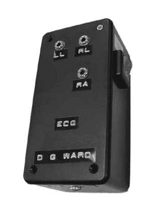
The preamplifier for ECG / EMG is identified as follows. For each set of measurements described below one electrode is plugged into LL and the other electrode is plugged into RA. A third, indifferent electrode, is plugged into RL. This provides for a lead II recording of ECG
Preparing the Subject
- Attach an electrode to the anterior surface of each forearm, about 2 to 3 in, above the wrist, and secure them with rubber straps. In the same manner, attach an electrode to each leg, approximately 2 to 3 in. above the medial malleolus (inner aspect of the ankle).
- Attach the appropriate tips of the patient cable to the electrodes. The cable leads are marked RA (right arm), LA (left arm), LL (left leg), and RL (right leg, the ground).
Sample ECG

Recording the ECG
The ECG will be recorded first under baseline (resting) conditions and then under conditions of fairly strenuous activity. Finally, recordings will be made while the subject holds his or her breath. The activity and breath holding recordings will be compared to the baseline recordings, and you will be asked to determine the reasons for the observed differences in the recordings.
BASELINE
- Position the subject comfortably in a supine position on a cot (if available), or sitting relaxed on a laboratory chair.
- Set the lead selector to the position corresponding to recording from lead I (RA-LA).
- Record the subject's ECG at-rest from lead II for 2 to 3 minutes or until the recording stabilizes. (You will need a tracing long enough to provide each student with a representative segment.) The subject should try to relax and not move unnecessarily, because the skeletal muscle action potentials will also be picked up and recorded.
"RUNNING IN PLACE"
- Make sure the electrodes are securely attached to prevent electrode movement while recording the EGG.
- Prepare to make the recording using lead II.
- Record the ECG while the subject is running in place for 3 min. Then have the subject sit down, but continue to record the ECG for an additional 4 minutes. Mark the recording at the end of the 3 minutes of running and at 1 minute after cessation of activity.
- Stop the recording.
"BREATH-HOLDING"
- Position the subject comfortably in the sitting position.
- Using lead II recording, begin the recording. After approximately 10 seconds, instruct the subject to begin breath holding and mark the record to indicate the onset of the 1-min breath-holding interval.
- Stop the recording after 1 minute and remind the subject to breathe.
Results:
BASELINE
- Identify and label the P, QRS, and T waves. Calculations you perform for your recording should be based on the following information: each large square represents an interval of 0.167 sec.
- Compute the heart rate. Determine the number of large
squares (and fraction thereof) from the beginning of one QRS complex to
the beginning of the next QRS complex, and plug this value into the
equation below to find the time for one heartbeat.
number of large squares x 0.167 sec = sec/beat
Now find the beats per minute, or heart rate, by using the figure just computed for the time for one heartbeat in the following equation:
Beats/min = 1/(sec/beat) x 60 sec/min = __________
Measure the QRS interval and compute its duration. __________
Measure the Q-T interval and compute its duration. __________
Measure the P-R interval and compute its duration. __________
- At the bottom of this page, attach segments of the ECG recordings from leads I through III. Make sure you indicate the timescale, lead, and subject's name on each tracing. To the recording on which you based your previous computations, add your calculations for the duration of the QRS, P-R intervals, and Q-T intervals above the respective area of tracing. Also record the heart rate on that tracing.
"RUNNING IN PLACE"
- Compute the beats/min during the third minute of running,
at 1 minute after exercise, and at 4 minutes after exercise. Record below:
Beats/min while running in place __________
Beats/min at 1 minute after exercise __________
Beats/min at 4 minutes after exercise. __________
"BREATH-HOLDING"
- Compute the beats/minute during the 1-minute experimental
(breath-holding) period.
Beats/min during breath holding __________
Discussion:
- Is the value obtained during baseline recording for heart rate within normal limits?
- Are the computed values during baseline recording for the QRS interval, the Q-T interval, the P-R interval within normal limits?
- Compare the recording during "running in place" with the baseline recording from lead II. Which wave intervals are shorter in the "running" recording? Does the subject's heart rate return to baseline levels by 4 minutes after exercise?
- If the cells of the SA node failed to function, what effect would this failure have on heart rate?
- Why is it important for impulses from the atria to be delayed at the AV node before they pass into the ventricles?
© David G. Ward, Ph.D. Last modified by wardd 23 May, 2006