Respiratory Control and acid-base balance
Objectives
- To define factors that influence the depth and rate of respiration.
- To examine the sounds made by air moving through the respiratory airways.
- To examine the role of carbonic acid - bicarbonate buffer system in stabilizing blood pH
Materials:
Group Supplies:
Tape measure
Nose clips
Alcohol swabs
70% ethanol solution
Disposable autoclave bag
Scotch tape
paper bag
Stethoscope
Pneumograph tubing
pressure - displacement transducer
pressure - displacement preamplifier (D. G Ward) (set to record activity within a range of DC to AC with variable gain)
digital to analog converter Dataq, DI-154)
computer with display software (WinDaq and WinDaq Browser)
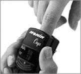
Nonin Pulse Oximeter
USE OF THE PNEUMOGRAPH TO DETERMINE FACTORS INFLUENCING RATE AND DEPTH OF RESPIRATION*
The neural centers that control respiratory rhythm and maintain a rate of 12 to 18 respirations/min are located in the medulla and pons. On occasion, input from the stretch receptors in the lungs (via the vagus nerve to the medulla) modifies the respiratory rate, as in cases of extreme over inflation of the lungs (Hering-Breuer reflex).
Death occurs when medullary centers are completely suppressed, as from an overdose of sleeping pills or gross overindulgence in alcohol, and respiration ceases completely.
Although the nervous system centers initiate the basic rhythm of breathing, there is no question that physical phenomena such as talking, yawning, coughing, and exercise can modify the rate and depth of respiration. So too can chemical factors such as changes in oxygen or carbon dioxide concentrations in the blood or fluctuations in blood pH. Changes in carbon dioxide blood levels seem to act directly on the medulla control centers, whereas changes in pH and oxygen concentrations are monitored by chemoreceptor regions in the aortic and carotid bodies, which in turn send input to the medulla. The experimental sequence in this section is designed to test the relative importance of various physical and chemical factors in the process of respiration.
The pneumograph, an apparatus that records variations in breathing patterns, is the best means of observing respiratory variations resulting from physical and chemical factors. The chest pneumograph is a coiled rubber hose that is attached around the thorax. As the subject breathes, chest movements produce pressure changes within the pneumograph that are transmitted to a recorder.
Methods and Results:
The instructor will demonstrate the method of setting up the pneumograph and discuss the interpretation of the results. Work in pairs so that one person can mark the record to identify the test for later interpretation. Ideally, the student being tested should face away from the recording apparatus to prevent voluntary modification of the record.
The amplifier for conditioning signals from the pneumograph is labeled pressure / displacement,
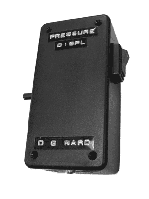
The pressure and displacement transducers are the following.
![]()
In addition to measuring respiration, measure heart rate and pO2 with the Nonin Pulse Oximeter.
- Attach the pneumograph tubing
firmly, but not restrictively, around the thoracic cage at the level of
the sixth rib, leaving room for chest expansion during testing. If the subject
is female, position the tubing above the breasts to prevent slippage
during testing. Record quiet breathing for 1 minute with the subject in a
sitting position.
Record breaths per minute. _______________________________ - Record a maximal inhalation followed
by a maximal exhalation. This should correlate to the vital capacity
measurement obtained in the spirometry lab, and will provide a baseline
for comparison during the rest of the pneumograph testing. Note the
direction the pneumograph recording moves during inspiration and during
expiration.
Measure in mm the height of the vital capacity recording. Divide the vital capacity recorded in the spirometry lab by the millimeter figure to obtain the volume (in milliliters of air) represented by one mm on the pneumograph recording. For example, if your vital capacity reading is 4000 mL and the vital capacity tracing occupies a vertical distance of 40 mm on the pneumograph recording, then a vertical distance of 1 mm equals 100 mL of air.
Record your computed value. ____________________ mL air/mm - Without recording, have the
subject breathe normally for 2 minutes, then inhale deeply and hold his or
her breath for as long as he or she can.
Time the breath-holding interval. _________________________ sec
As the subject exhales, turn on the recording apparatus and record the recovery period (time to return to normal breathing — usually slightly over 1 minute):
Time of recovery period. ______________________________ sec
Did the subject have the urge to inspire or expire during breath holding?
________________________________________________________________
Without recording, exhale completely and forcefully after taking a deep breath. What was observed this time? How long could the subject hold their breath?
______________________________________________________________
______________________________________________________________
Explain the results. (Hint: the vagus nerve is the sensory nerve of the lungs and plays a role here.)
___________________________________________________________
___________________________________________________________ - Have the subject
hyperventilate (breathe deeply and forcefully at the rate of 1 breath/4
sec) for about 30 seconds.
Caution: A sensation of dizziness may develop. (As the carbon dioxide is washed out of the blood by overventilation, the blood pH increases, leading to a decrease in blood pressure and reduced cerebral circulation.) The subject may experience a lack of desire to breathe after forced breathing is stopped. If the period of breathing cessation — apnea-is extended, cyanosis of the lips may occur.
Record both during and after hyperventilation. How does the pattern obtained during hyperventilation compare with that recorded during the vital capacity tracing?
____________________________________________________________
Is the respiratory rate after hyperventilation faster or slower than during normal quiet breathing?
______________________________________________________________ - Repeat the above test, but do not record until after hyperventilating. After hyperventilation, the subject is to hold his or her breath as long as he or she can. Can the breath be held for a longer or shorter period of time after hyperventilating?
- Without recording, have the
subject breathe into a paper bag for 3 minutes, then record his or her
breathing movements.
Caution: During the bag-breathing exercise the subject's partner should watch the subject carefully for any untoward reactions.
Is the breathing rate faster or slower than that recorded during normal quiet breathing?
__________________________________________________________________
After hyperventilating?_______________________________________ - Run in place for 2 minutes,
and then have your partner determine the length of time that you can hold
your breath.
Length of breath-holding. _____________________ Sec - To prove that respiration has
a marked effect on circulation, conduct the following test. Have your lab
partner record the rate and relative force of your radial pulse before
beginning.
rate _______________ beats/min relative force __________________
Inspire forcibly. Immediately close your mouth and nose to retain the inhaled air, and then make a forceful and prolonged expiration. Your lab partner should observe and record the condition of the blood vessels of your neck and face, and again immediately palpate the radial pulse.
Observations ___________________________________________
____________________________________________________________
Radial pulse ____________________ beats/min Relative force _______________
Explain the changes observed. _________________________________________ - Dispose of the paper bag in the autoclave bag.
11. Record the subject's breathing as he or she performs activities from the list below. Make sure the record is marked accurately to identify each test conducted
|
talking |
swallowing water |
|
yawning |
coughing |
|
laughing |
lying down |
|
standing |
running in place |
|
doing a math problem |
|
© David G. Ward, Ph.D. Last modified by wardd 23 May, 2006